PET camera reveals body mechanisms
Professor Lauri Nummenmaa’s four-year project at the University of Turku is using groundbreaking technology to study the connections between anxiety and heart symptoms.
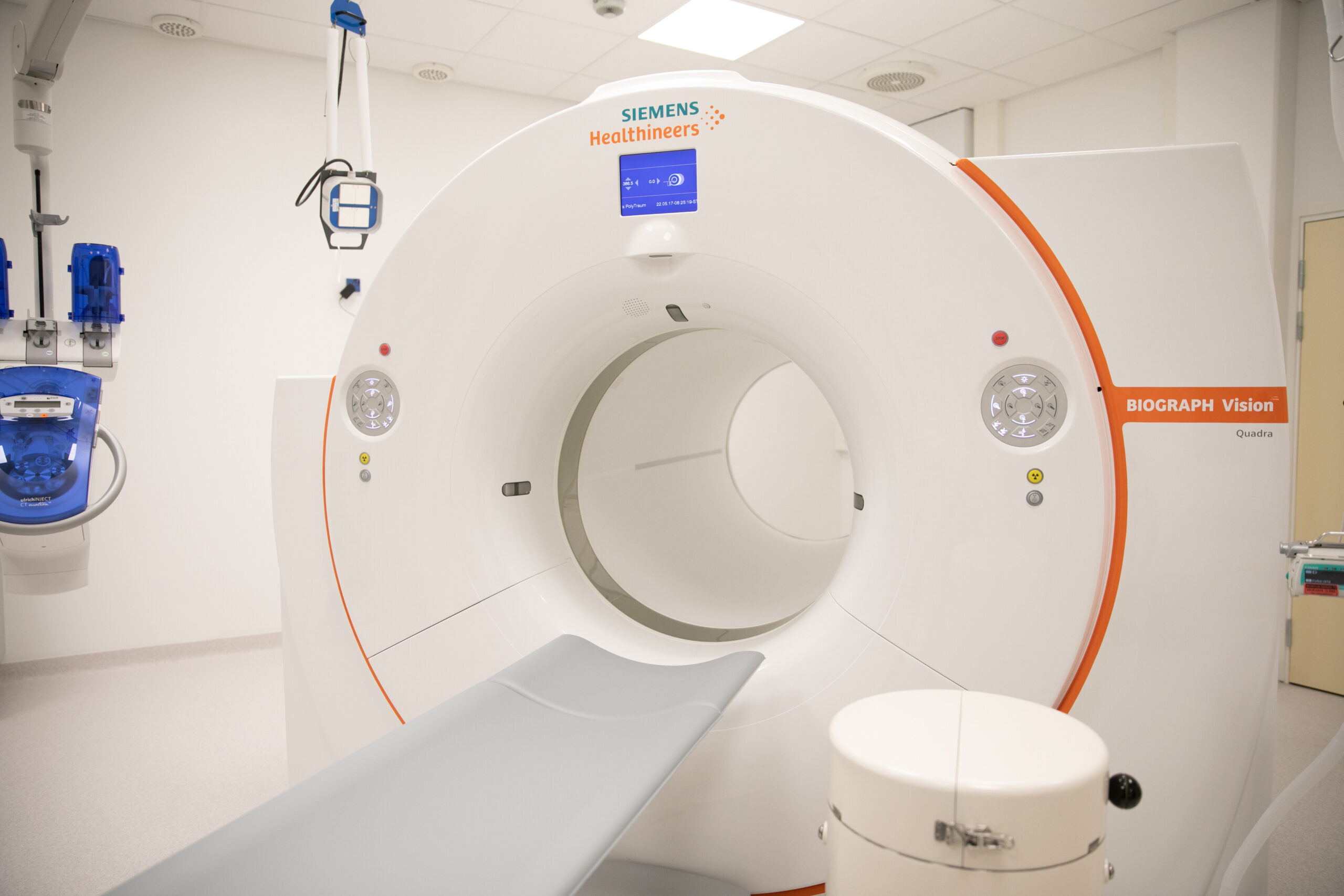
PET camera captures a person from head to thigh in one go from a distance of just over a metre.
(Photo: University of Turku)
When we fall in love, our hearts pound. When we are nervous, our stomach churns. When we are frightened, a cold shiver runs down our spine.
The mind and body are often perceived as being separate from each other, but the reality of humanity is not so dichotomous. Even emotions don’t just arise in our brain, they change how the whole body works. The phenomenon has been known in psychology for a long time. In medicine, too, attention has been paid to, for example, how various mental illnesses show symptoms in the body.
Obtaining accurate information on the topic has been difficult. Although brain imaging has long shown how sensations activate different areas of the brain, the rest of the body has remained in the shade. In practice, the connections between body and mind have been mapped, for example, through studies in which people have been asked to describe how sensations affect the body.
However, new technologies create new opportunities. This is the case with the new generation PET camera, or positron emission tomography camera, acquired for Turku PET Centre in 2022.
The camera captures a person from head to thigh in one go from a distance of just over a metre.
“This enables us to see in real time how emotional reactions occur simultaneously in both the brain and the body,” Nummenmaa says.
Radioactive water shows emotions throughout the body
In March, Nummenmaa and his team received a grant of €720,000 from the Jane and Aatos Erkko Foundation for a project that aims to collect comprehensive information on how heart diseases and anxiety symptoms appear throughout the body.
“We know that there is a link between anxiety disorders and heart symptoms,” Nummenmaa explains.
“Anxiety can cause chest pain, and on the other hand, coronary artery disease, for example, is often accompanied by anxiety disorders. The new PET camera enables us to see in real time how, for example, the anxiety response activates in different parts of the brain, heart, blood circulation and other organs.”
In practice, this is done using radioactive water, where ordinary oxygen has been replaced by a radioactive oxygen-15 isotope. Water is administered into the bloodstream to see how blood flow changes in different parts of the body.
“Blood flow indicates the activity of organs and the body. If a lot of blood flows somewhere, it means that things are happening there.” After isotope administration, the test subject can, for example, be shown a frightening film while a PET camera monitors the movements of radioactive water in the bloodstream. This provides real-time information on where the body reacts to anxiety and where it does not.
The isotope has a half-life of just two minutes and allows imaging to take about four minutes. The test subject receives about as much radiation as during a flight across the Atlantic.
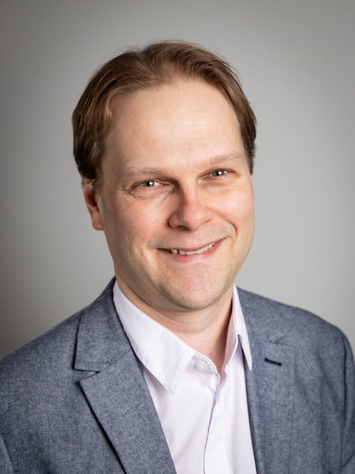
Lauri Nummenmaa
Professor, PET Centre, University of Turku
Group leader in the project Imaging the link between anxiety and cardiovascular diseases using total-body positron emission tomography funded by the Jane and Aatos Erkko Foundation.
(Photo: University of Turku)
In the verge of something completely new
This is a completely new kind of basic research, Nummenmaa says. Since there are only a few full-body PET cameras, no one else is doing similar research.
During the four-year study, he hopes to photograph hundreds of research subjects. With the help of repetitions, our perception of how the body works will become more accurate.
“If we find clear signs of, for example, factors threatening an anxiety disorder and their presence in the scan, this would be a great help in diagnosing anxiety.”
In addition to radioactive water, Nummenmaa is also planning imaging using fluorine-labelled glucose. This can be used to accurately measure the body’s energy consumption.
“Energy consumption gives us a slightly different perspective on the same things.”
However, the circulation of sugar is slower than that of water and cannot be monitored in the same way in real time. This makes it suitable for mapping the effects of long-term anxiety or heart symptoms.
The goal is to prevent depression
But why such a gloomy research topic? Instead of anxiety, wouldn’t it be more fun to study, say, the effects of joy on the body?
“Researchers naturally have the freedom to study what they want, but freedom also comes with responsibility,” Nummenmaa replies.
He points out that anxiety disorders are a major public health problem, affecting up to 7% of Finns. Diseases of the cardiovascular system, on the other hand, are the most common cause of death in Finland. It is the researcher’s duty to conduct research that promotes well-being and innovations.
“The purpose of the research is to better understand these diseases and their mechanisms. When we learn to understand the signs of anxiety, for example, we can develop treatments that will hopefully prevent anxiety proactively.”
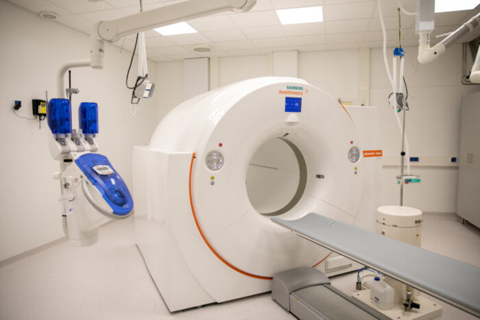
There are only a few full-body PET cameras in the world. In 2021 the Jane and Aatos Erkko Foundation awarded a grant of €5 million for the purchase of the PET camera to PET Centre of the University of Turku.
(Photo: University of Turku)
Full-body PET revolutionised research
Headed by Nummenmaa, the research is one of a couple of dozen new research projects launched after the acquisition of a new camera at the Turku PET Centre, a joint national research institute of the University of Turku, Turku University Hospital and Åbo Akademi University.
Professor Juhani Knuuti, Head Physician at the Centre, says the camera has revolutionised research work throughout the Centre
“Whereas the old camera was usually used to photograph one organ at a time, you can now see almost the entire body at the same time. It has made it possible to look at things holistically,” Knuuti describes.
At the same time, this has required new kinds of research groups.
“For example, I myself am a cardiologist. Now, however, I can see that cardiac events affect the brain or intestines, for example, but I don’t understand anything about them. So I need people in my group who do.”
There are a total of around a hundred researchers using the PET camera.
Exceptional grant
The purchase of the PET camera was an expensive €7.5 million investment. The Jane and Aatos Erkko Foundation awarded a grant of €5 million for it in 2021.
The grant is a very exceptional one, as the Foundation does not, as a rule, support equipment purchases. However, the importance of the PET camera was supported by its national usability and significance for the extensive development of research and diagnostics.
There are only a few similar devices in the world.
Turku has been particularly enthusiastic about research carried out with radioactive water. Their topics range from emotions and heart disease to issues such as obesity, metabolism and brown fat.
About half of those imaged are research subjects. The other half is clinical work of the hospital.
“But clinical patients, too, are imaged throughout. Only clinically requested points are used in medical reports, but everything else is good research material,” Knuuti sums up.